CNS: Practice Question
Question
What is the most likely diagnosis of this cytology specimen from the CNS?
Question images
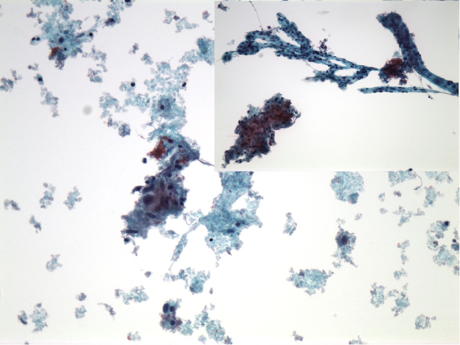
CNS cytology specimen with an inset showing an additional microscopic field.
Answer Choices
- Glioblastoma multiforme
- Metastatic clear cell renal cell carcinoma
- Metastatic seminoma
- Oligodendroglioma
- Classic ependymoma
Show Answer and Explanations
Glioblastoma multiforme — Correct answer
Correct. Glioblastoma multiforme shows markedly pleomorphic cells, necrosis that may form pseudopalisading on cell block and vascular proliferation. The central image shows markedly pleomorphic cells and necrosis, and the image at the upper right highlights vascular proliferation.
Metastatic clear cell renal cell carcinoma
Incorrect. Clear cell renal cell carcinoma shows monolayered cells with foamy vacuolated cytoplasm, eccentrically placed nuclei with small nucleoli and minimal atypia.
Metastatic seminoma
Incorrect. Seminoma shows large mostly single cells with fine chromatin and a central nucleolus, with a background of lymphocytes and a ‘tigroid’ pattern.
Oligodendroglioma
Incorrect. Oligodendroglioma shows more monotonous nuclei and may have a granular background.
Classic ependymoma
Incorrect. A classic ependymoma shows enlarged epithelioid cells, with finely granular cytoplasm, eccentrically located nuclei with small nucleoli and may show rosettes and pseudorosettes.
Summary
Summary: Glioblastoma multiforme shows markedly pleomorphic cells, necrosis that may form pseudopalisading on cell block and vascular proliferation.Free account required. No subscription needed.