CSF: Practice Question
Question
Which is the most likely diagnosis from this cytology specimen from the CSF?
Question images
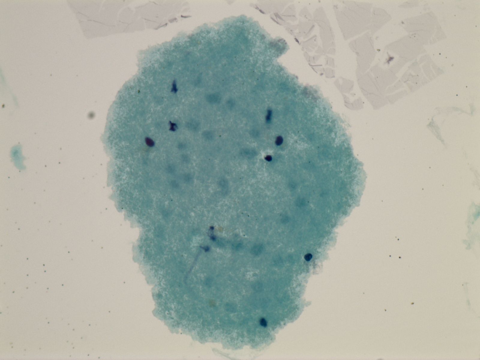
Cerebrospinal fluid cytology specimen.
Answer Choices
- Normal tissue
- Anaplastic large cell lymphoma
- Metastatic adenocarcinoma
- Metastatic squamous cell carcinoma
- Metastatic melanoma
Show Answer and Explanations
Normal tissue — Correct answer
Correct. This is normal brain tissue, and is more likely to be seen in the CSF after procedures or with the presence of foreign objects such as shunts.
Anaplastic large cell lymphoma
Incorrect. Anaplastic large cell lymphoma shows large pleomorphic cells. The cytoplasm is basophilic and vacuolated. The nuclei are round, with prominent nucleoli and clumpy chromatin. It is positive for T cell markers such as CD2, CD3 or CD5, positive for CD30, and is divided into ALK positive (better prognosis) and ALK negative (worse prognosis).
Metastatic adenocarcinoma
Incorrect. Adenocarcinoma shows three dimensional clusters of malignant cells frequently with vacuoles and prominent nucleoli.
Metastatic squamous cell carcinoma
Incorrect. Metastatic squamous cell carcinoma shows hyperchromatic angular nuclei, may show keratinization and abnormal forms such as "tadpole" forms.
Metastatic melanoma
Incorrect. Metastatic melanoma shows single malignant cells with very prominent nucleoli and may show intracytoplasmic melanin.
Summary
Summary: Normal brain tissue is more likely to be seen in the CSF after procedures or with the presence of foreign objects such as shunts.Free account required. No subscription needed.